Background: Calcifying odontogenic cyst is a developmental cyst that often appears in jaw cysts, and dentigerous cyst is the most common developmental cyst of the jaw, which has the ability to turn into a neoplasm. p53 is increased in some odontogenic cysts and tumors. The aim of this study is to compare the expression of P53 marker in dentigerous cyst and calcifying odontogenic cyst by immunohistochemical method. Methods: In this descriptive-analytic cross-sectional study, the studied population is the sample of odontogenic dentigerous cysts and calcifying odontogenic cysts in the pathology center of urmia. First, all the files in the archive of the department from 2000 to 2021 were examined and the cases of cysts were examined separately and in terms of the expression of P53 marker in dentigerous cysts and calcifying odontogenic cysts by immunohistochemical method. Results: The results showed that the overall mean expression of p-53 marker in dentigerous cyst group was 33.45 with a standard deviation of 11.86%, among which the lowest and highest expression of the said marker was 12% and 54%, respectively. The overall average expression of p-53 marker in the calcifying odontogenic cyst group is 59.55 with a standard deviation of 13.73%, among which the lowest and highest expression of the said marker was 30% and 87%, respectively. There is a statistically significant difference between dentigerous cyst patients and odontogenic calcifying cyst patients in terms of p-53 marker expression, and the said marker is more expressed in patients in the odontogenic calcifying cyst group compared to dentigerous cyst patients (p< 0.001) Conclusions: It seems that this protein (p53) can probably be a suitable marker to confirm the ability to predict invasion in premalignant oral lesions such as cysts in odontogenic calcification patients.
This is an Open Access article, distributed under the terms of the Creative Commons Attribution 4.0 International License (http://creativecommons.org/licenses/by/4.0/), which permits unrestricted use, distribution and reproduction in any medium or format, provided the original work is properly cited.
. This cyst was first described by Rywkind in 1932
[2]
Rajkumar K, Kamal K, Sathish M, Leena S. Calcifying odontogenic cyst. Journal of Oral and Maxillofacial Pathology. 2004; 8(2): 99.
[2]
. The calcifying odontogenic cyst has significant variation in clinical features, histopathology and also biological behavior, and several classifications have been proposed for this lesion
[1]
Kamboj M, Juneja M. Ameloblastomatous Gorlin' s cyst. Journal of oral science. 2007; 49(4): 319-23.
Toida M. So‐called calcifying odontogenic cyst: review and discussion on the terminology and classification. Journal of oral pathology & medicine. 1998; 27(2): 49-52.
. In most cases, a cyst appears to be benign, while in some cases, it does not have any characteristics of a cyst, which may be tumoral
[5]
Seyedmajidi M, Nafarzadeh S, Siadati S, Shafaee S, Bijani A, Keshmiri N. p53 and PCNA expression in keratocystic odontogenic tumors compared with selected odontogenic cysts. International journal of molecular and cellular medicine. 2013; 2(4): 185. URL:
. In 2005, the World Health Organization described cystic calcifying odontogenic cyst type as a low-grade malignancy and changed its name to calcifying cystic odontogenic tumor and introduced its solid type as dentinogenic gum cell tumor, which is a very aggressive nature. has
[6]
AHN SG, KIM SA, KIM SG, LEE SH, Kim J, YOON JH. β‐catenin gene alterations in a variety of so‐called calcifying odontogenic cysts. Apmis. 2008; 116(3): 206-11.
Dentigerous cyst is the most common facial developmental cyst
[8]
Fatemeh M, Sepideh A, Sara BS, Nazanin M. P53 protein expression in dental follicle, dentigerous cyst, odontogenic keratocyst, and inflammatory subtypes of cysts: An immunohistochemical study. Oman medical journal. 2017; 32(3): 227.
, which occurs in most cases in the second and third decade of life and is mostly related to the third molar of the mandible and the canine of the maxilla
[9]
Bernick S. Dentigerous cysts of the jaw. Oral Surgery, Oral Medicine, Oral Pathology. 1949; 2(7): 914-21.
Knights E, Brokaw W, Kessler H. The incidence of dentigerous cysts associated with a random sampling of unerupted third molars. Gen Dent. 1991; 39(2): 96-8.
[9, 10]
. This cyst is clinically asymptomatic, but it has the potential to become massively enlarged and cause cortical expansion and erosion
[11]
Daley TD, Wysocki GP. The small dentigerous cyst: a diagnostic dilemma. Oral Surgery, Oral Medicine, Oral Pathology, Oral Radiology, and Endodontology. 1995; 79(1): 77-81.
Shafer WG. A text book of oral pathology. Cherubism. 1983: 699-702.
[11, 12]
. The dentigerous cyst is reduced through the hydrostatic pressure caused by the accumulation of fluid between the enamel epithelium. and an unerupted tooth crown is created
[13]
Veera SD, Padanad G. Dentigerous cyst with recurrent maxillary sinusitis: a case report with literature review. Int J Appl Dent Sci. 2015; 1(4): 16-9.
[14]
Wang C-J, Huang P-H, Wang Y-L, Shyng Y-C, Kao W-B. Dentigerous cyst over maxillary sinus: A case report and literature review. 2009; 20(2): 116-24.
[13, 14]
. This cyst sometimes turns into squamous cell carcinoma, mucoepidermoid carcinoma or ameloblastoma
[15]
Khambete N, Kumar R, Risbud M, Kale L, Sodhi S. Dentigerous cyst associated with an impacted mesiodens: report of 2 cases. Imaging Science in Dentistry. 2012; 42(4): 255-60.
P53 protein is the product of a tumor suppressor gene called TP53
[5]
Seyedmajidi M, Nafarzadeh S, Siadati S, Shafaee S, Bijani A, Keshmiri N. p53 and PCNA expression in keratocystic odontogenic tumors compared with selected odontogenic cysts. International journal of molecular and cellular medicine. 2013; 2(4): 185. URL:
. The half-life of P53 is short and about 20 minutes
[5]
Seyedmajidi M, Nafarzadeh S, Siadati S, Shafaee S, Bijani A, Keshmiri N. p53 and PCNA expression in keratocystic odontogenic tumors compared with selected odontogenic cysts. International journal of molecular and cellular medicine. 2013; 2(4): 185. URL:
. Under normal conditions, this protein is continuously produced and in the nucleus of the cell, it is connected to MDM2 protein (mouse double minute 2) until this complex is transported to the cytoplasm, where it is degraded by proteosomes. This process keeps the cellular concentration of P53 low
[16]
Gaballah ETM, Tawfik MA. Immunohistochemical analysis of P53 protein in odontogenic cysts. The Saudi Dental Journal. 2010; 22(4): 167-70.
. Therefore, increasing the concentration of this protein depends on inhibiting its degradation, under stress, certain types of protein are released from the nucleus to the nucleoplasm and prevent the exit of P53 to the cytoplasm and Subsequently, it prevents its decomposition and this protein accumulates in the nucleus
[17]
Olson M, Dundr M. The moving parts of the nucleolus. Histochemistry and cell biology. 2005; 123(3): 203-16.
. Considering that the production amount of P53 is low and its half-life is short, it is possible to detect it in conditions where the protein is expressed in high amounts or accumulated inside mutated cells
[18]
Ayla Ö, Can T, Mehmet Y, Belir A, Gülçin E. Expression of the tumor suppressor gene p53 in odontogenic cysts. Balkan Journal of Stomatology. 2002; 6(1): 47-50.
[19]
Oliveira MGd, Lauxen IdS, Chaves ACM, Rados PV, Sant' Ana Filho M. Immunohistochemical analysis of the patterns of p53 and PCNA expression in odontogenic cystic lesions. Medicina oral, patología oral y cirugía bucal Valencia Vol 13, no 5 (May 2008), p e275-e280. 2008.
[18, 19]
.
Immunohistochemical studies regarding the expression of P53 in odontogenic cysts have obtained conflicting results
[16]
Gaballah ETM, Tawfik MA. Immunohistochemical analysis of P53 protein in odontogenic cysts. The Saudi Dental Journal. 2010; 22(4): 167-70.
Kichi E, Enokiya Y, Muramatsu T, Hashimoto S, Inoue T, Abiko Y, et al. Cell proliferation, apoptosis and apoptosis‐related factors in odontogenic keratocysts and in dentigerous cysts. Journal of oral pathology & medicine. 2005; 34(5): 280-6.
. Therefore, this study will be conducted in order to find a relationship between cystic changes and P53 expression, taking into account the difference in the clinical behavior of these two cysts, which are common developmental cysts, until the expression level of this protein in the said cysts is determined and with Compare each other.
2. Material and Method
The sample size is based on the study of Setara Shojaei et al.
[23]
Shojaee S, Jamshidi S, Mohtasham N, Roshanaee G, Shahabinejad M. Evaluation of MDM2 and P53 Expression in Dentigerous Cyst and Odontogenic Keratocyst by Immunuhistochemistry. Journal of Mashhad Dental School. 2015; 39(2): 163-72.
and considering the statistical power of 80%, the second type error of 20%, and the confidence level of 85%. According to the mentioned study, the parameters including the mean and standard deviation of P53 expression in calcifying odontogenic cyst groups were 36.03 ± 17.28 and the same mean in dentigerous cyst was 25.51 ± 11.76. The sample size for each group was estimated to be 11 samples, and due to conducting the study in the form of separated groups and considering the drop in the number of samples, in order to increase the power of the study and detect differences, we took the sample size of 20 samples in each group from the beginning.
The present study was carried out using an analytical method. The studied population is the samples of odontogenic cysts, dentigerous cells and calcifying odontogenic cysts in the pathology department of the specialized pathology laboratory. First, all the files in the archive of the department from 2012 to 2022 were examined and the cases of cysts were separated. The H&E slides of these cases were studied and the histopathological diagnosis will be confirmed based on the existing criteria. All cases of non-inflammatory cysts were selected.
Considering things such as the presence of sufficient tissue in the paraffin block, the thickness and sufficient amount of epithelium in the sample stained with H&E, the presence of minimal bleeding, and the presence of information related to background variables (age, sex, and location of the lesion), Selected cases were prepared for immunohistochemical staining.
From the paraffin blocks of each sample, two sections with a thickness of 5 microns were prepared and placed on a glass slide prepared with polylysine. The standard method of STREPTAVIDIN-IMMUNOPEROXIDASE BIOTIN was used for staining.
First, the prepared sections will be deparaffinized by xylene and dehydrated using diluted alcohol solutions. Endogenous peroxidase activity will be inhibited by incubating the samples with 2% peroxydehydrogen in methanol for thirty minutes. For antigen retrieval, the samples were microwaved for 15 minutes in 0.01 M citrate buffer and pH 6, at a high degree.
After that, washing was done for three consecutive times for 5 minutes each time with tris buffer sulfate. To avoid non-specific reactions, the sections were incubated with 10% serum for 10 minutes. Then the reaction with p53 antibody (prediluted, Copenhagen, Dako, Denmark) was performed for one hour at room temperature.
In the next step, the samples will be washed with PBS in three times of 5 minutes. The reaction with the secondary antibody will be done at room temperature for 30 minutes and washing with PBS was done again. After that, the samples were incubated with DAB for 5-10 minutes and stained with Mayer's hematoxylin as a counterstain.
The positive control for p53 marker was a positive sample of squamous cell carcinoma of the tongue and the negative control was by substituting non-reactive mouse serum instead of the primary antibody. Sections stained with p53 protein were observed under a light microscope and brown colored cells were considered as positive. Cell counting was done with x400 magnification and the ratio of stained cells to total cells was measured. Based on this, the amount of stained cells is 0-5% as negative, 25-5% as +, 25-50% was considered as ++, more than 50% as +++.
The data was coded in spss software version 20 and analyzed at a significant level of 0.05 using standard statistical tests. Quantitative demographic data with average and standard deviation and qualitative demographic data with frequency and percentage in the form of tables and in cases where necessary; It was presented as a diagram for better understanding. The normality of quantitative data was evaluated using the Kolmogro-Smirnoff test, and according to the distribution of normality and non-normality of quantitative variables, standard parametric and non-parametric tests were selected and applied. To compare the average occurrence of the gene under investigation in the groups, if the distribution was normal, the t-test was used, and otherwise, the non-parametric Mann-Whitney test was used. In comparison of these averages according to age groups, if normal, analysis of variance test was used, otherwise Kruskal-Wallis test was used. Chi-score test is used to compare the frequency of clinical information according to gender, and to evaluate the relationship between quantitative variables in the groups under investigation, Pearson's correlation coefficient was used if it was normal, and Spearman's correlation coefficient was used otherwise.
3. Results
In the present study, the overall average expression of p-53 marker in the calcifying odontogenic cyst group was 59.55 with a standard deviation of 13.73%, among which the lowest and highest expression of the said marker was 30% and 87%, respectively. the overall average expression of p-53 marker in the dentigerous cyst group was 33.45 with a standard deviation of 11.86%, among which the lowest and highest expression of the said marker was 12 and 54%, respectively.
Table 1. Distribution of frequency and percentage of gender between the two groups.
Cyst type
men
women
total number (percent)
number
percent
number
percent
Dentigerous cyst
8
40
12
60
20 (100)
calcifying odontogenic cyst
10
50
10
50
20 (100)
Dentigerous cyst and calcifying group Odontogenic cysts have been somewhat uniform in terms of gender distribution.
Table 2. Overall average expression percentage of p-53 marker in people with odontogenic cysts under investigation In the present study.
overall average
Standard deviation
lowest
highest
The percentage of p53 marker expression
46.50
18.30
12
87
The overall average expression of p-53 marker in people with odontogenic cysts under investigation is 46.50 with a standard deviation of 18.30%, among which the lowest and highest expression of the said marker was 12% and 87%, respectively.
Table 3. Overall mean percentage of p-53 marker expression in the group.
overall average
Standard deviation
lowest
highest
The percentage of p53 marker expression
33.45
11.86
12
54
The overall average expression of p-53 marker in the dentigerous cyst group was 33.45 with a standard deviation of 11.86%, among which the lowest and highest expression of the said marker was 12 and 54%, respectively.
Table 4. The overall average expression percentage of p-53 marker in the calcifying odontogenic cyst group.
overall average
Standard deviation
lowest
highest
The percentage of p53 marker expression
59.55
13.73
30
87
The overall mean expression of p-53 marker in the calcifying odontogenic cyst group was 59.55 with a standard deviation of 13.73%, among which the lowest and highest expression of the said marker was 30% and 87%, respectively.
Table 5. Comparison of p-53 marker expression percentage by gender in the present study.
men
women
P-value
overall average
Standard deviation
overall average
Standard deviation
The percentage of p53 marker expression
47.22
18.61
45.91
18.47
0.001<
The data in the above table compares the mean expression percentage of p-53 marker by gender in the present study by using the t-test, and its data shows that there is a statistical difference between the mean expression percentage of p-53 marker in men and women. There is significance and the said marker is more expressed in men than in women (p=<0.001).
Table 6. Comparison of the expression percentage of p-53 marker according to the groups under investigation in the present study.
dentigerous cyst
calcifying odontogenic cyst
P-value
overall average
Standard deviation
overall average
Standard deviation
The percentage of p53 marker expression
33.45
11.86
59.55
13.73
0.001<
Explanation: The data in the above table are compared using the t-test to compare the average expression percentage p-53 marker in two groups under investigation in this study and its data shows that there is a statistically significant difference between dentigerous cyst patients and odontogenic calcifying cyst patients in terms of p-53 marker expression and the said marker in Patients in the calcifying odontogenic cyst group are more expressed compared to patients in the dentigerous cyst group (p<0.001). (Tables 1-5)
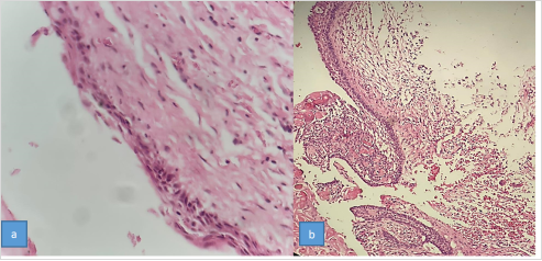
Microscopic views of H&E and IHC staining (DC&COC) are presented in Figures 1 and 2.
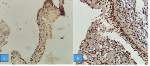
Figure 2. P53 Immunohistochemistry staining (IHC) in the groups: a (dentigerous cyst), b (calcifying odontogenic cyst) × 10 magnification.
4. Discussion
Odontogenic cysts and tumors are an important part of jaw and oral lesions that arise from remnants of odontogenic epithelium in different stages of odontogenesis
[24]
Neville BW, Damm DD, Allen C, Chi AC. Oral and maxillofacial pathology: Elsevier Health Sciences; 2015.
[24]
, in the present study, 20 people have dentigerous cysts and 20 people have calcifying odontogenic cysts. The expression of p-53 marker has been investigated and compared, and the results of the present study in terms of the number of samples compared to other comparable studies such as Shojaei et al.'s study
[23]
Shojaee S, Jamshidi S, Mohtasham N, Roshanaee G, Shahabinejad M. Evaluation of MDM2 and P53 Expression in Dentigerous Cyst and Odontogenic Keratocyst by Immunuhistochemistry. Journal of Mashhad Dental School. 2015; 39(2): 163-72.
KHalili M. Immunohistochemical investigation of p53 and ki67 proteins in samples of inflammatory and developmental odontogenic cysts. Iranian Journal of Dentistry. 2020; 22(3): 182-9.
[25]
The sample is higher, so it has the required validity. These cases are considered a strength in the generalizability of the results of this study.
In the present study, 45% of men and 22% of women in both dentigerous cyst and calcifying odontogenic cyst groups have been examined and compared in terms of p-53 marker expression. This means that both dentigerous cyst and calcifying odontogenic cyst groups were the same in terms of gender distribution (Table 1). Therefore, the gender variable as a potential confounding variable cannot bias the results. This case also indicates the validity of the results of the present study, away from potential confounding variables that may underreport or overreport the results.
The findings showed that in the present study, the overall average expression of the p-53 marker in the subjects with odontogenic cysts under investigation was 46.50 with a standard deviation of 18.30%, among which the lowest and highest expression of the said marker was 12% and 87%, respectively (Table 2). Also, the findings indicated that in the current study, the overall average expression of the p-53 marker in the dentigerous cyst group was 33.45 with a standard deviation of 11.86%, among which the lowest and highest expression of the said marker was 12% and 54%, respectively (Table 3). In the present study, the overall mean expression of p-53 marker in the calcifying odontogenic cyst group was 59.55 with a standard deviation of 13.73%, among which the lowest and highest expression of the said marker was 30% and 87%, respectively (Table 4).
In previous studies such as the study of Gurgel et al.
[26]
Gurgel CAS, Ramos EAG, Azevedo RA, Sarmento VA, da Silva Carvalho AM, dos Santos JN. Expression of Ki-67, p53 and p63 proteins in keratocyst odontogenic tumours: an immunohistochemical study. Journal of molecular Histology. 2008; 39(3): 311-6.
AHN SG, KIM SA, KIM SG, LEE SH, Kim J, YOON JH. β‐catenin gene alterations in a variety of so‐called calcifying odontogenic cysts. Apmis. 2008; 116(3): 206-11.
in this regard, the expression of p53 marker was reported between 41 and 91 percent. Perhaps one of the reasons for this variation in the occurrence of the mentioned marker is the number of sample sizes, different study design types, and differences in measurement tools. One of the other reasons for justifying these conflicting results in the complex biology of P53 is access to antibody, the effect of tissue preparation and the protocol used for immunohistochemistry, such as fixation methods, antigen retrieval method from paraffin blocks, and the type of antibody. Both the stated reasons are effective in the accuracy of the results and the variation in the percentage of occurrence of p53 marker in different cysts.
The findings showed that there is a statistically significant difference between the mean expression percentage of p-53 marker in women and men, and the said marker is expressed more in men than in women (Table 5). This finding was previously reported in the study of Abou-Bakr et al.
[27]
Abou-Bakr AA, Abdelaziz AA, Malash IA, Mansour O, Abdelsalam IM, Abo-Elazm OM, et al. The Prognostic Significance of c-Met and p53 Immunohistochemical Expression in Gastric and Colorectal Carcinomas. Open Access Macedonian Journal of Medical Sciences. 2021; 9(A): 134-42.
Zydroń R, Marszałek A, Bodnar M, Kosikowski P, Greczka G, Wierzbicka M. The analysis of expression of p16 protein in group of 53 patients treated for sinonasal inverted papilloma. Brazilian Journal of Otorhinolaryngology. 2018; 84: 338-43.
The findings also showed that there is a statistically significant difference between dentigerous cyst patients and calcifying odontogenic cyst patients in terms of p-53 marker expression, and the said marker is more in patients in the calcifying odontogenic cyst group compared to patients in the dentigerous cyst group. It is stated (Table 6). This finding was consistent with the study by Shujaei et al.
[23]
Shojaee S, Jamshidi S, Mohtasham N, Roshanaee G, Shahabinejad M. Evaluation of MDM2 and P53 Expression in Dentigerous Cyst and Odontogenic Keratocyst by Immunuhistochemistry. Journal of Mashhad Dental School. 2015; 39(2): 163-72.
. These results indicate that the occurrence of p53 marker in these two groups can indicate the biological behavior of these two cysts. One of the other reasons for the occurrence of this difference in the two groups and the lower incidence of p53 marker in the dentition in the dentigerous cyst compared to the calcifying odontogenic cyst may be the fact that, on the other hand, the main trigger for the formation of dentigerous cysts is not clearly known; But often an inflammatory infiltration is observed in the capsule of the cyst, which causes epithelial cells to multiply early. This problem (response to inflammatory stimulation) may cause the growth process of the cyst, but the proliferation of cells may be non-continuous and only in short periods of time, which probably causes a decrease in the expression of P53 in dentigerous cysts. Therefore, the statement Less p53 in dentigerous cysts could be due to cellular stress produced by inflammatory stimuli, although this cyst.
It has an evolutionary origin
[29]
Kadashetti V, Patil N, Datkhile K, Kanetakar S, Shivakumar K. Analysis of expression of p53, p63 and proliferating cell nuclear antigen proteins in odontogenic keratocyst: An immunohistochemical study. Journal of Oral and Maxillofacial Pathology: JOMFP. 2020; 24(2): 273.
Gadbail A, Chaudhary M, Patil S, Gawande M. Actual Proliferating Index and p53 protein expression as prognostic marker in odontogenic cysts. Oral Diseases. 2009; 15(7): 490-8.
, a lower incidence of p53 marker was reported in dentigerous cyst compared to odontogenic calcifying cyst. P53 gene mutation can be associated with increased cell proliferation
[26]
Gurgel CAS, Ramos EAG, Azevedo RA, Sarmento VA, da Silva Carvalho AM, dos Santos JN. Expression of Ki-67, p53 and p63 proteins in keratocyst odontogenic tumours: an immunohistochemical study. Journal of molecular Histology. 2008; 39(3): 311-6.
. The high expression of this protein in the epithelial lining of odontogenic calcifying cyst may mean more proliferative activity and as a result, more aggressive biological behavior of this lesion compared to dentigerous cyst
[31]
Barreras CMU, Camou JGR, Burgueño ERR. P63/p40 expression in benign odontogenic cysts and tumors. Revista Odontológica Mexicana. 2022; 25(3): 215-23.
The findings indicate that there is a statistically significant difference between dentigerous cyst patients and odontogenic calcifying cyst patients in terms of p-53 marker expression and the said marker in patients in the odontogenic calcifying cyst group compared to the dentigerous cyst group patients It is further stated; So this protein can probably be a suitable marker to confirm the ability to predict invasion in oral premalignant lesions such as cysts occurring in odontogenic calcification patients.
Acknowledgments
This article is an exception from the dissertation of the General Doctor of Dentistry with the code of ethics IR.UMSU.REC.1400.197. We would like to thank Urmia University of medical sciences for supporting us in this research.
Author Contributions
Samira Mostafazadeh: Conceptualization, Project administration, Supervision, Writing – original draft
Rajkumar K, Kamal K, Sathish M, Leena S. Calcifying odontogenic cyst. Journal of Oral and Maxillofacial Pathology. 2004; 8(2): 99.
[3]
Toida M. So‐called calcifying odontogenic cyst: review and discussion on the terminology and classification. Journal of oral pathology & medicine. 1998; 27(2): 49-52.
Seyedmajidi M, Nafarzadeh S, Siadati S, Shafaee S, Bijani A, Keshmiri N. p53 and PCNA expression in keratocystic odontogenic tumors compared with selected odontogenic cysts. International journal of molecular and cellular medicine. 2013; 2(4): 185. URL:
AHN SG, KIM SA, KIM SG, LEE SH, Kim J, YOON JH. β‐catenin gene alterations in a variety of so‐called calcifying odontogenic cysts. Apmis. 2008; 116(3): 206-11.
Fatemeh M, Sepideh A, Sara BS, Nazanin M. P53 protein expression in dental follicle, dentigerous cyst, odontogenic keratocyst, and inflammatory subtypes of cysts: An immunohistochemical study. Oman medical journal. 2017; 32(3): 227.
Knights E, Brokaw W, Kessler H. The incidence of dentigerous cysts associated with a random sampling of unerupted third molars. Gen Dent. 1991; 39(2): 96-8.
[11]
Daley TD, Wysocki GP. The small dentigerous cyst: a diagnostic dilemma. Oral Surgery, Oral Medicine, Oral Pathology, Oral Radiology, and Endodontology. 1995; 79(1): 77-81.
Shafer WG. A text book of oral pathology. Cherubism. 1983: 699-702.
[13]
Veera SD, Padanad G. Dentigerous cyst with recurrent maxillary sinusitis: a case report with literature review. Int J Appl Dent Sci. 2015; 1(4): 16-9.
[14]
Wang C-J, Huang P-H, Wang Y-L, Shyng Y-C, Kao W-B. Dentigerous cyst over maxillary sinus: A case report and literature review. 2009; 20(2): 116-24.
[15]
Khambete N, Kumar R, Risbud M, Kale L, Sodhi S. Dentigerous cyst associated with an impacted mesiodens: report of 2 cases. Imaging Science in Dentistry. 2012; 42(4): 255-60.
Ayla Ö, Can T, Mehmet Y, Belir A, Gülçin E. Expression of the tumor suppressor gene p53 in odontogenic cysts. Balkan Journal of Stomatology. 2002; 6(1): 47-50.
[19]
Oliveira MGd, Lauxen IdS, Chaves ACM, Rados PV, Sant' Ana Filho M. Immunohistochemical analysis of the patterns of p53 and PCNA expression in odontogenic cystic lesions. Medicina oral, patología oral y cirugía bucal Valencia Vol 13, no 5 (May 2008), p e275-e280. 2008.
[20]
Suzuki T, Kumamoto H, Kunimori K, Ooya K. Immunohistochemical analysis of apoptosis‐related factors in lining epithelium of radicular cysts. Journal of oral pathology & medicine. 2005; 34(1): 46-52.
Kichi E, Enokiya Y, Muramatsu T, Hashimoto S, Inoue T, Abiko Y, et al. Cell proliferation, apoptosis and apoptosis‐related factors in odontogenic keratocysts and in dentigerous cysts. Journal of oral pathology & medicine. 2005; 34(5): 280-6.
Shojaee S, Jamshidi S, Mohtasham N, Roshanaee G, Shahabinejad M. Evaluation of MDM2 and P53 Expression in Dentigerous Cyst and Odontogenic Keratocyst by Immunuhistochemistry. Journal of Mashhad Dental School. 2015; 39(2): 163-72.
Neville BW, Damm DD, Allen C, Chi AC. Oral and maxillofacial pathology: Elsevier Health Sciences; 2015.
[25]
KHalili M. Immunohistochemical investigation of p53 and ki67 proteins in samples of inflammatory and developmental odontogenic cysts. Iranian Journal of Dentistry. 2020; 22(3): 182-9.
[26]
Gurgel CAS, Ramos EAG, Azevedo RA, Sarmento VA, da Silva Carvalho AM, dos Santos JN. Expression of Ki-67, p53 and p63 proteins in keratocyst odontogenic tumours: an immunohistochemical study. Journal of molecular Histology. 2008; 39(3): 311-6.
Abou-Bakr AA, Abdelaziz AA, Malash IA, Mansour O, Abdelsalam IM, Abo-Elazm OM, et al. The Prognostic Significance of c-Met and p53 Immunohistochemical Expression in Gastric and Colorectal Carcinomas. Open Access Macedonian Journal of Medical Sciences. 2021; 9(A): 134-42.
Zydroń R, Marszałek A, Bodnar M, Kosikowski P, Greczka G, Wierzbicka M. The analysis of expression of p16 protein in group of 53 patients treated for sinonasal inverted papilloma. Brazilian Journal of Otorhinolaryngology. 2018; 84: 338-43.
Kadashetti V, Patil N, Datkhile K, Kanetakar S, Shivakumar K. Analysis of expression of p53, p63 and proliferating cell nuclear antigen proteins in odontogenic keratocyst: An immunohistochemical study. Journal of Oral and Maxillofacial Pathology: JOMFP. 2020; 24(2): 273.
Gadbail A, Chaudhary M, Patil S, Gawande M. Actual Proliferating Index and p53 protein expression as prognostic marker in odontogenic cysts. Oral Diseases. 2009; 15(7): 490-8.
@article{10.11648/j.sf.20250604.12,
  author = {Samira Mostafazadeh and Babak Ebrahimi and Fariba Abdal},
  title = {Comparison of P53 Marker Expression Between Dentigerous Cyst and Calcifying Odontogenic Cyst},
  journal = {Science Frontiers},
  volume = {6},
  number = {4},
  pages = {133-139},
  doi = {10.11648/j.sf.20250604.12},
  url = {https://doi.org/10.11648/j.sf.20250604.12},
  eprint = {https://article.sciencepublishinggroup.com/pdf/10.11648.j.sf.20250604.12},
  abstract = {Background: Calcifying odontogenic cyst is a developmental cyst that often appears in jaw cysts, and dentigerous cyst is the most common developmental cyst of the jaw, which has the ability to turn into a neoplasm. p53 is increased in some odontogenic cysts and tumors. The aim of this study is to compare the expression of P53 marker in dentigerous cyst and calcifying odontogenic cyst by immunohistochemical method. Methods: In this descriptive-analytic cross-sectional study, the studied population is the sample of odontogenic dentigerous cysts and calcifying odontogenic cysts in the pathology center of urmia. First, all the files in the archive of the department from 2000 to 2021 were examined and the cases of cysts were examined separately and in terms of the expression of P53 marker in dentigerous cysts and calcifying odontogenic cysts by immunohistochemical method. Results: The results showed that the overall mean expression of p-53 marker in dentigerous cyst group was 33.45 with a standard deviation of 11.86%, among which the lowest and highest expression of the said marker was 12% and 54%, respectively. The overall average expression of p-53 marker in the calcifying odontogenic cyst group is 59.55 with a standard deviation of 13.73%, among which the lowest and highest expression of the said marker was 30% and 87%, respectively. There is a statistically significant difference between dentigerous cyst patients and odontogenic calcifying cyst patients in terms of p-53 marker expression, and the said marker is more expressed in patients in the odontogenic calcifying cyst group compared to dentigerous cyst patients (p< 0.001) Conclusions: It seems that this protein (p53) can probably be a suitable marker to confirm the ability to predict invasion in premalignant oral lesions such as cysts in odontogenic calcification patients.},
 year = {2025}
}
TY  - JOUR
T1  - Comparison of P53 Marker Expression Between Dentigerous Cyst and Calcifying Odontogenic Cyst
AU  - Samira Mostafazadeh
AU  - Babak Ebrahimi
AU  - Fariba Abdal
Y1  - 2025/12/09
PY  - 2025
N1  - https://doi.org/10.11648/j.sf.20250604.12
DO  - 10.11648/j.sf.20250604.12
T2  - Science Frontiers
JF  - Science Frontiers
JO  - Science Frontiers
SP  - 133
EP  - 139
PB  - Science Publishing Group
SN  - 2994-7030
UR  - https://doi.org/10.11648/j.sf.20250604.12
AB  - Background: Calcifying odontogenic cyst is a developmental cyst that often appears in jaw cysts, and dentigerous cyst is the most common developmental cyst of the jaw, which has the ability to turn into a neoplasm. p53 is increased in some odontogenic cysts and tumors. The aim of this study is to compare the expression of P53 marker in dentigerous cyst and calcifying odontogenic cyst by immunohistochemical method. Methods: In this descriptive-analytic cross-sectional study, the studied population is the sample of odontogenic dentigerous cysts and calcifying odontogenic cysts in the pathology center of urmia. First, all the files in the archive of the department from 2000 to 2021 were examined and the cases of cysts were examined separately and in terms of the expression of P53 marker in dentigerous cysts and calcifying odontogenic cysts by immunohistochemical method. Results: The results showed that the overall mean expression of p-53 marker in dentigerous cyst group was 33.45 with a standard deviation of 11.86%, among which the lowest and highest expression of the said marker was 12% and 54%, respectively. The overall average expression of p-53 marker in the calcifying odontogenic cyst group is 59.55 with a standard deviation of 13.73%, among which the lowest and highest expression of the said marker was 30% and 87%, respectively. There is a statistically significant difference between dentigerous cyst patients and odontogenic calcifying cyst patients in terms of p-53 marker expression, and the said marker is more expressed in patients in the odontogenic calcifying cyst group compared to dentigerous cyst patients (p< 0.001) Conclusions: It seems that this protein (p53) can probably be a suitable marker to confirm the ability to predict invasion in premalignant oral lesions such as cysts in odontogenic calcification patients.
VL  - 6
IS  - 4
ER  -